articles

articles

articles

articles


Our eyes are extremely delicate, yet they can be subjected to harsh conditions and other environmental factors that affect their health. One of the problems that can affect our eyes is an accumulation of dirt, debris and bacteria on the eyelids. This can cause a range of issues, including stopping tear film from reaching the eyes and being properly dispersed over their surface – which is necessary to keep them healthy and comfortable. Fortunately, a new solution called Blephex can help.
What is Blephex?
Blephex is a handheld electro-mechanical device that is applied to the margins of the eyelids with the purpose of cleaning them and improving the effectiveness with which tear film flows onto the surface of the eyes.
Blephex has a disposable, surgical-grade sponge tip which rapidly oscillates to create a cleaning action. Before the sponge tip is placed onto the eyes, it is soaked in a gentle exfoliating solution. This solution provides soft abrasion to help remove dead skin cells and debris that could be irritating the eyes and interrupting tear film progression. The BlephexÔ device is manually applied to the eyes and moved gently across the eyelids, with the entire, painless process taking approximately 6 to 8 minutes per eye. A different sponge is used on each eye, ensuring that no bacteria is passed between them. After the procedure, patients are given instructions on how to maintain the cleanliness of their eyelids with daily/nightly eyelid hygiene at home.
Most patients experience a significant improvement in tear film production and dispersal, and a reduction in unpleasant symptoms that they may have been experiencing within 48 hours of their treatment. While a single treatment is normally enough to produce excellent results, many patients are advised to have Blephex every 4-6 months.
What conditions can Blephex help with?
Blephex can be used to clean the eyelids at any time, and people who suffer from dry eyes or eye allergies may find it is particularly beneficial for helping to reduce the symptoms that they experience. It can also be combined with Lipiflow – another technological solution – to help counteract the effects of dry eyes.
Unsurprisingly, Blephex is particularly recommended as a treatment for an eye condition called blepharitis. Blepharitis is characterized by the inflammation of the eyelids, which causes them to become red, swollen and itchy. Although the condition is not usually serious, it can lead to further problems if it isn’t treated.
Symptoms of blepharitis include:
- Sore eyes
- Itchy eyes
- A gritty, irritated feeling affecting the eyes
- Redness
- Flakes or crustiness around the roots of the eyelashes
- Eyelids that stick together when you wake up in the morning
If you are suffering from the symptoms of blepharitis, dry eyes or eye allergies and feel that you would benefit from Blephex treatment, please contact our team to schedule a consultation appointment.

Neurolens are the first and only prescription lenses that include an element of contoured prism in their design. This prism is designed to bring the patient’s eyes into more equal alignment, and this should help to provide relief from the symptoms that are associated with several eye misalignment conditions, including digital eye strain and binocular vision dysfunction.
What is digital eye strain?
Digital eye strain is the name given to describe a group of symptoms that can occur when someone spends long periods of time using digital devices. Since using digital devices requires the eyes to work harder than normal and we don’t always position our devices the perfect distance away, it can lead to issues such as eye pain, dry and irritated eyes, eye fatigue, light sensitivity and blurred vision. Unsurprisingly, the number of people who are experiencing digital eye strain has grown significantly over the last few years and is expected to continue to do so.
What is binocular vision dysfunction?
Binocular vision dysfunction, also known as BVD for short, is another eye condition but is one that is very misunderstood. Binocular vision dysfunction occurs when the eyes aren’t perfectly aligned, causing your brain and eyes to work harder than normal in order to create a clear visual image and remain focused. This places pressure on the trigeminal nerve, which is the nerve that is responsible for the majority of the sensations that we experience in our head and back. BVD can often manifest as other things owing to the huge range of symptoms that are associated with the condition. These can include, but aren’t limited to:
- Blurred vision
- Headaches/migraines
- Double vision
- Motion sickness
- Vertigo
- Dizziness
- Anxiety
Many people don’t think to visit an eye doctor when they are experiencing these symptoms, but all can occur simply because the eyes are out of alignment.
What are Neurolens lenses and how do they help?
As well as containing your usual prescription, Neurolens lenses also contain a specific amount of contoured micro-prism. This micro-prism alters the position of images so that they are aligned in the same plane. This then reduces the pressure on the muscles around the eyes as well as bringing the eyes into alignment, easing the symptoms that the patient has been experiencing.
The amount of prism in Neurolens lenses is decided using the Neurolens eye-tracking device. This non-invasively measures the misalignment that the patient is experiencing, and this is used to form the basis for the patient’s Neurolens prescription. After this, it’s fairly normal for the amount of prism to need to be adjusted by infinitesimal amounts to achieve the optimal relief from your symptoms. Most patients who choose Neurolens treatment see a 50% improvement in their vision as soon as they start to have micro-prism incorporated into their prescription lenses. However, with careful adjustments, many patients see as much as an 80% reduction in the effects of digital eye strain and binocular vision dysfunction.
Want more information about Neurolens? Please contact our knowledgeable eye care specialists.

Visian ICL, or “implantable Collamer lens” is an alternative procedure for patients who may not be ideal candidates for Lasik or other alternative corrective eye surgery. Visian ICL is typically used for patients who do not want to remove portions of their cornea, have thin corneas, or that have excessively high levels of nearsightedness (myopia).
This procedure makes a small incision and then implants a personalized prescription lens over the cornea to allow for corrected vision. If your vision then changes due to aging or other natural processes, the lens can be replaced by another lens with an updated prescription.
Who is a Good Candidate for ICL?
Typically, patients that would benefit from ICL are between the ages of 21-45. This age represents a slight increase from the base age of 18 for Lasik. This procedure is also not well suited for geriatric or elderly individuals. Patients may also have mild or severe myopia, and they have a prescription that has been relatively unchanged. While the age requirements are more stringent for ICL than Lasik, there are other less stringent qualifications. This means that even if you aren’t an ideal candidate for Lasik, ICL could be a good option for you.
Performing the Operation
ICL is considered an outpatient operation and only takes about 30 minutes to complete. This means that you will be in and out of your chosen facility on the same day. Patients are given some numbing drops for their eyes and individuals that are more hesitant or uncomfortable may also be given a sedative.
The surgeon will make several micro-incisions in the eye to insert and place the lens. When the lens has been inserted, it will be unfolded, and the edges of the lens will be placed behind the iris. After this is completed, the operation is considered complete. Your physician may give you some eye drops for postoperative care and then send you home. There may be a follow-up appointment scheduled 24 hours later.
Post-Operation
After your operation, you will be required to have somebody else drive you home. Anytime that you have an operation that may impair your vision or ability to operate a vehicle, you should plan to have somebody else drive you home. Surgery results are typically noticeable 24 hours after the operation.
Recovery time is minimal, and some patients experience mild discomfort or a gritty feeling in their eyes. Your doctor may require you to stay out of the swimming pool and avoid activities that make you heavily perspire because when sweat gets into your eyes, it may aggravate the micro incisions and cause additional discomfort.
While there are some potential complications both during and after the operation, they are typically minimal. The chances of impairing your vision or causing long-term damage are very low with this operation, however, you should make sure to talk about potential side effects with your physician. If you experience any abnormality, you should seek medical attention immediately.

Dry eyes are one of the most common conditions that can affect our eyes and is estimated to affect millions of Americans. As you’ve probably guessed, dry eyes occur when tears fail to provide enough natural lubrication for the eyes to be comfortable and healthy. Exactly what causes dry eyes can vary significantly, from side effects from medications to prolonged computer use. What is clear is that while the condition isn’t sight-threatening, it can make day to day life much harder than it needs to be. Fortunately, there are treatments that can help, and arguably one of the most effective is Lipiflow.
What is Lipiflow?
Lipiflow is a new technological solution that addresses the underlying cause of your dry eyes, rather than simply treating the symptoms. It is most effective at helping patients whose dry eyes are caused by meibomian gland dysfunction – a condition characterized by problems with the way that the meibomian glands produce the oil that forms an essential part of our tear film. The meibomian glands can become less productive, or in some cases, even blocked by hardened oil deposits. This prevents the oil from reaching your tear film, making it less effective. Lipiflow targets the meibomian glands, warming them to break down oily blockages and massaging your eyes to make sure that the oil, and then the tear film, is evenly dispersed. This helps to combat the symptoms associated with dry eyes, which can include:
- Eye fatigue
- Dry, scratchy and uncomfortable eyes
- Blurred vision
- Sensitivity to light
- Difficulty wearing contact lenses
Your eye doctor will be able to advise you if Lipiflow has the potential to be a suitable solution for your dry eyes.
What to expect from Lipiflow treatment?
Lipiflow treatment is a simple, painless process that is performed in the comfort of your eye doctor’s office. There is no need for anesthetic. Once you are settled in your chair, your eye doctor will open the sterile, single-use applicators which are placed over your eyes. These are connected to a machine that causes the inner eyelids to heat to approximately 42.5°C to, while simultaneously placing gentle pressure on the outer eyelid surfaces. Lipiflow takes around 12 minutes per eye, during which time you can relax. You can even listen to music if you’d like to. There is no downtime, and patients can return to their usual activities right away. It takes around 3 days for patients to begin to see an improvement in their dry eye symptoms, although they may require further treatment in the future to maintain them. Optimal results are usually achieved around 6 to 8 weeks following your Lipiflow treatment.
For more information about Lipiflow, or to schedule a consultation to talk about this treatment for dry eyes, please contact our office.

LASIK (laser-assisted in situ keratomileusis), is the most popular refractive surgical procedure. In this procedure, a laser is used to permanently change the shape of the cornea (the clear covering on the front of the eye) to correct common vision problems such as nearsightedness, farsightedness, astigmatism, and presbyopia. This improves vision and reduces a person's need for glasses or contact lenses.
LASIK uses an excimer laser (an ultraviolet laser) to remove a thin layer of corneal tissue, giving the cornea a new shape, so that light rays are focused clearly on the retina. In the case of a nearsighted person, the goal of LASIK is to flatten the too-steep cornea; with farsighted people, a steeper cornea is desired. LASIK can also correct astigmatism by smoothing an irregular cornea into a more normal shape.
LASIK is an outpatient surgical procedure with no need to stay at the surgery center overnight as it will take 10 to 15 minutes to perform for each eye. The procedure is done while the patient is awake, but the patient may request mild sedation. The only anesthetic used is eye drops that numb the surface of the eye. LASIK can be done on one or both eyes during the same session.
How to Prepare for LASIK Eye Surgery?
Before LASIK eye surgery, the eye surgeon will evaluate the patient’s medical history and perform a full eye examination, including measuring corneal thickness, refraction, corneal mapping, eye pressure, and pupil dilation. Afterward, the surgeon will discuss what to expect during and after the procedure.
On the day of the surgery, eat a light meal before going to the doctor and take all prescribed medications, if any. Do not wear eye makeup, creams, perfumes or lotions on the day before and the day of surgery, or have any bulky hair accessories that will interfere with positioning head under the laser.
Contact lenses shouldn't be worn for at least three days prior to the evaluation. In the case of, rigid gas permeable contact lenses, they should not be worn for at least three weeks before. Patients should arrange for a ride home from the place of surgery, as their vision might be blurry.
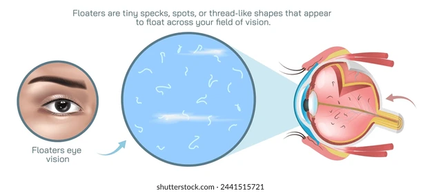
What are floaters?
Eye floaters are those small spots, threads, or cobweb-like shapes that float around in your field of vision, especially when looking at a bright, plain background like the sky or a white wall. They’re often more noticeable when you're looking at something bright or when your surroundings are uniform, and they move as you move your eyes.
Floaters are usually harmless and are often caused by age-related changes in the vitreous humor, the gel-like substance that fills the inside of your eye. As we age, the vitreous can shrink and become more liquid, causing tiny fibers to clump together and cast shadows on the retina, which you perceive as floaters.
However, if you notice a sudden increase in floaters, especially if they’re accompanied by flashes of light or a loss of peripheral vision, it’s important to see an eye doctor. This could be a sign of a more serious condition like a retinal tear or detachment.
Causes of Eye Floaters
Aging (Most Common Cause)
As we get older, the vitreous humor (the gel-like substance that fills the eye) can shrink and become more liquid. This change causes tiny fibers within the vitreous to clump together, casting shadows on the retina, which we see as floaters.Retinal Detachment or Tear
A more serious cause of floaters can be a retinal detachment or tear. When the vitreous shrinks, it can sometimes tug on the retina, leading to a tear or detachment. This can cause a sudden increase in floaters, along with flashes of light. If left untreated, this can lead to permanent vision loss.Inflammation of the Eye (Uveitis)
Inflammation inside the eye, especially in the middle layer (the uvea), can cause floaters. Conditions like uveitis can release cells into the vitreous, creating floaters.Diabetic Retinopathy
High blood sugar levels from diabetes can damage the blood vessels in the retina, leading to leakage of blood or fluid into the vitreous. This can result in floaters.Eye Injury or Surgery
Any trauma to the eye or surgery (like cataract surgery) can cause floaters, either from physical changes to the eye or from bleeding into the vitreous.Myopia (Nearsightedness)
People with myopia are at a higher risk for floaters. The elongated shape of the eyeball in myopia can cause the vitreous to separate from the retina more easily.Medication Side Effects
Some medications, such as those used for treating certain eye conditions (like intravitreal injections), may cause changes in the vitreous and result in floaters.Posterior Vitreous Detachment (PVD)
This is a natural age-related process where the vitreous humor pulls away from the retina. It can lead to floaters, and though it’s typically harmless, it can sometimes lead to complications.
Symptoms
Spots or Shapes
You might see spots, squiggly lines, or cobweb-like shapes that seem to float in your line of sight. These are the floaters themselves. They might appear darker or lighter depending on your background.Movement with Eye Movement
Floaters usually move when you move your eyes. They seem to "drift" in the direction you’re looking, often staying in your peripheral vision.Increased Floaters
If you notice a sudden increase in the number of floaters or if they appear more intensely, it might indicate something more serious, like a retinal tear or detachment.Flashes of Light
Flashes of light, also known as photopsia, may accompany the floaters if there’s tension on the retina or a retinal tear. These are often described as lightning-like flashes or sudden bursts of light.Blurred Vision
In some cases, especially with more severe eye conditions like retinal detachment, you might experience blurred vision in addition to floaters and flashes.Loss of Peripheral Vision
If a retinal detachment is present, floaters may be accompanied by a gradual loss of side vision or a "curtain" effect that may obscure parts of your vision.
When Should You See a Doctor?
If you notice any of the following symptoms, it's important to seek immediate medical attention:
A sudden increase in floaters.
Flashes of light, especially if they’re new.
A shadow or "curtain" over part of your vision.
Sudden vision loss.
Even though most floaters are harmless and common with aging, these warning signs could point to a retinal tear, detachment, or other serious eye condition, and early intervention is key to preventing vision loss.

Eyelid rejuvenation surgery is a medical procedure that is designed to reduce the appearance of bagginess from the lower eyelids and sagging from the upper eyelids. This operation is often used for cosmetic surgery to reduce the appearance of aging.
This surgery can also be considered when the eyelids are interfering with a patient’s ability to see. Sometimes a sagging upper eyelid can partially obscure the eyeball, interfere with a person’s ability to look in certain directions or interfere with their peripheral vision.
Why Do My Eyelids Sag?
As our skin ages, it often loses its elasticity. This doesn’t allow the skin to rebound back to its original shape and we see the appearance of wrinkles and bulges that aren’t due to weight gain. While these wrinkles can occur all over the body, they typically first appear in the face and eyes. Any additional skin on or near the eyelids can cause the eyelid to sag or droop over the eyelashes and into the frame of vision.
The eyelids also contain some fat to protect and cushion the eyeball. The fat is held in place by a thin membrane. As we age, the membrane can weaken and will no longer keep the fat in place. This fat can create bulges in the upper and lower eyelids.
What Can I Do About Eyelid Sag or Bulging?
There are several non-surgical treatments on the market to help treat sagging skin or reduce the appearance of wrinkles, but it is important to use additional caution when applying any product near the eyes. Always make sure that the product is designed to be used in the predetermined area. If you have questions about a specific product, you should contact your health care provider.
Many individuals find that non-surgical options don’t have enough or any effect on their eyelids. In these instances, they can consider working with a licensed medical professional to weigh their options. Eyelid rejuvenation (blepharoplasty) is also commonly called an eye lift.
There are several different methods for achieving your desired results. Typically, an incision is made into the eyelid, and then the excess skin or fat cells are removed by laser or scalpel. Additionally, your surgeon may suggest that you also get laser resurfacing done in combination with your surgery.
What to Expect
Eyelid surgery can be done in a local office or a surgery center. If you are completing the operation in an office environment, you can probably expect that you will be treated with a local anesthetic and an oral sedative. If you are in a surgery center, it’s likely that you will receive an intravenous anesthetic. The surgery takes about two hours to complete if you are getting all four eyelids done (upper and lower lids). When you are getting all four eyelids corrected, the surgeon will likely opt to work on both upper eyelids first and then move to the lower eyelids. While the upper eyelids will have three to six stitches, the lower eyelids may not have any. The stitches should remain in place for three to six days.

We all want to enjoy healthy eyes and clear vision for as long as we possibly can. Fortunately, there are things that we can do to help make this possible. It’s easy to think that protecting our eyes from obvious harm, such as the harsh effects of UV light or potential injury during an activity like welding or woodworking, is enough. However, there are many other things that we can do to keep our eyes functioning well for as long as possible. This includes taking supplements to help ensure that we get the right nutrition for our eyes to be healthy.
EyePromise offers a range of highly effective and expert-recommended nutritional supplements that are designed to support optimal eye health.

Eye disease that is caused by diabetes is currently the number one cause of blindness and vision loss. Due to the increased risk in diabetic patients, doctors recommend that people over 30 with diabetes get an annual dilated eye exam. Diabetic patients under 30 should get this exam five years after they have been diagnosed.
Diabetic retinopathy is a condition that is caused by damage to the retina. Patients that have diabetes may also have experienced extended periods of time where their blood sugar was elevated. The high levels of blood sugar damage the retina’s walls which leave them susceptible to leaking. When fluid accumulates in the retina or macula, it causes vision loss.
To make these matters worse, if prolonged high blood sugar levels are seen again, the retina will be oxygen-depleted. This causes the abnormal growth of new blood vessels. This condition is called neovascularization. This blood vessel type is weak and prone to leaking. As these blood vessels leak, they introduce blood into the eye. Excessive bleeding into the eye can cause blindness.
Treatment
While a healthy diet and exercise can be beneficial to your optical health, diabetic retinopathy is a condition that is caused by damage to the retinal wall. While this damage can sometimes be corrected, simple diet changes won’t reverse the effects.
It is essential to catch the condition in the earlier stages to reduce the effects. This can also help patients understand the importance of monitoring their blood sugar so that repeat events can be limited. Treatment options are even more successful when diabetic retinopathy is caught early. These options include vitrectomy, scatter photocoagulation and focal photocoagulation.
During both scatter, and focal photocoagulation the doctor will use lasers to help alleviate the condition. The lasers make small burns on the retina aimed at the blood vessels. These burns will help to seal the blood vessels to prevent more leakage and stop them from growing larger.
When using scatter photocoagulation, hundreds of small burns are made in a specific pattern during two additional appointments. Scatter coagulation should be used on patients who do not have advanced diabetic retinopathy.
Focal photocoagulation specifically targets the leaking blood vessels that are in the macula. Unfortunately, this procedure is not aimed to correct the blurry vision associated with diabetic retinopathy, but it does stop it from progressing further. Once the retina has detached, neither form of photocoagulation can be used.
Vitrectomy is a surgery that helps to remove scar tissue and/or the fluid that is clouded with blood that has been leaked into the eye. This operation is the most successful when performed before the disease has progressed too far. When the operation only targets removing the fluid, success rates are very high for the procedure. When the procedure also aims to reattach the retina, the failure rate is around 50%.

Premium IOLs or intraocular lenses are lenses that are placed in the eye during cataract surgery. The lens placement is designed to restore the natural lens shape. These lenses can also be placed as a vision correction device called refractive lens exchange. Premium IOLs offer advanced features beyond the single vision IOL’s that are also offered. These features include aspheric, toric, accommodating, and multifocal IOL’s.
Understanding Premium IOL Types
Aspheric Lenses
These lenses closely match the natural curve of the eye. Typical lenses were uniformly curved making it easier to manufacture, but at the same time increasing the chance of causing imperfections in vision. Aspheric lenses help to reduce imperfections and improve clarity, especially at nighttime.
Toric IOLs
These lenses are specifically designed to help correct nearsightedness, farsightedness, and astigmatism.
Accommodating IOLs
Accommodating IOL’s can tilt slightly forward when you look at objects that are close to the eye. This helps to improve visibility when you are performing actions like reading a book. While they are not necessarily as sharp as bifocals, patients have a reduced need to use reading glasses while still maintaining excellent distance vision.
Multifocal IOLs
If you require a bifocal or trifocal lens in your glasses, this may be a likely choice for you. Different portions of the lens allow for better vision at different ranges. However, there are some overall sacrifices with vision clarity at a distance.
History of Premium IOLs
Premium IOLs have been approved by the Food and Drug Administration (FDA) since the 1980s. Prior to FDA approval, when patients had cataract surgery, they were required to wear very thick eyeglasses or specialized contact lenses to correct their vision. New technologies in the optical world have allowed for a wide variety of available premium IOLs and figuring out which specific type that suits you best will depend on some different factors.
Patient Factors
Physicians are careful to discuss the realities of this procedure with their patients. After recovering from cataract surgery, many patients expect that their vision will be completely restored to their peak performance. However, doctors are careful to warn against this and explain the realities of the surgery and as well as likely expectations of what will result from the surgery. For this reason, surgeons are likely to have some initial concern about the desired outcome for the patient to make sure that their hopes are grounded.
Surgeons will also have an eye toward the patient's desire to not wear eyeglasses. If the patient does not mind wearing corrective lenses without the need for surgery, this may be the best option. These lenses may also not be an ideal fit for the elderly population. Eyes in the geriatric population are often rapidly deteriorating requiring a lens replacement more quickly than would be recommended.
Patients with certain medical histories may also be poor candidates for premium IOL surgery. Some of these conditions include:
Advanced macular degeneration
Anterior basement membrane dystrophy
Fuch’s dystrophy
Weak zonules
Glaucoma
Post-refractive surgery patients
This list is not comprehensive, so it’s important to consult with your physician and bring a detailed medical history for their review.
Finally, patients may also want to consider their careers when weighing the value of this surgery. Patients who are required to read on computer screens for extended periods of time (i.e., print editors, office jobs) may be ideal candidates.
In contrast, individuals that require long-distance acuity like truck drivers, pilots, or even photographers may find that some of the issues with these lenses are not suited to their needs. Individuals often complain of “halos” during the night when looking toward a light, glare, or general acuity issues at longer distances.
Further Consideration
While premium IOLs do have some limitations, they offer an excellent choice for many individuals. However, it is important to meet with your eye care professional to fully discuss all of the available options to find your best fit as well as to make sure that you understand all of the potential risks and restrictions that this operation poses.


Office Hours
Mon: 12:00 PM to 6:00 PM
Tue: 10:00 AM to 4:00 PM
Wed: 10:30 AM to 4:00 PM
Thu: 10:00 AM to 4:00 PM
Fri - Sun: Closed
Doctor's Hours
Mon: 12:30 PM - 6:00 PM
Tue: 10:00 PM - 4:00 PM
Wed: 11:30 AM - 4:00 PM
Thu: 10:00 PM - 4:00 PM
Powered by 